 We have available in our laboratory two custom-built anterior segment optical coherence tomography systems. The first one is a time-domain OCT system (primarily used for in vitro applications) and a spectral-domain OCT system (built in collaboration with Copernicus University in Torun, and used in vivo).
 The time-domain system was built in a free air Michelson interferometer configuration. The low
coherence light source was a SLD with a central wavelength of 820nm and 20nm of bandwidth. The light coming from the SLD was collimated by a pigtailed lens. The reference arm consists of a stepper motor configured with a speed of 23.75mm/s, which provides a central Doppler frequency of 58.6 kHz. In the sample arm, a galvanometer scanner is used together with plano-convex lens aperture as a collimation lens for chief rays. Finally, the light returned by the sample and reference arm is collected by a fiber optic pigtail, which drives the light to a balanced avalanche photodiode detector (Hamamatsu, Hamamatsu, Japan). The OCT signal is then filtered by a custom bandpass filter of central frequency of 58. 6 kHz and bandwidth of 2.4 and digitized by a PCI card and stored in the computer to produce the images. A very useful feature of this setup is its capability to capture confocal images of the sample without any additional element. Taking advantage of the speed of the galvanometric scanner (1 kHz) it is possible to use this feature for almost a real-time preview (about 2 frames per second with satisfying resolution of 400 × 400 pixels) of the sample. (Ortiz et al. 2009b)
The spectral domain system is based on fiber-optics Michelson interferometer configuration with a superluminescent diode SLD (central wavelength of 840 nm, and 50 nm bandwidth) serving as a source and a 12-bit line-scan CMOS camera with 4096 pixels serving as a detector. The horizontal and vertical scanning was made with use of two galvanometer optical scanners. The axial resolution of this system was measured to be about 3.4 microns in air. A high acquisition rate, up to 135,000 A-scans/second enables three-dimensional reconstruction of the anterior segment during lenticular accommodation,
blinking and pupillary reaction to light stimulus. In this system, the axial range is doubled (to up to 10 mm) using a complex conjugate image removal (capture of time and spectral domain signals). (Grulwoski et al. 2009)
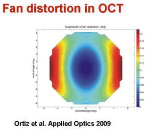
The anterior segment images obtained from OCT are subject to fan and optical distortion. We have developed techniques for hardware optimization and software compensation of fan distortion, which arises from the fan distortion architecture (Ortiz et al. 2009b). In addition, we have developed algorithms based on 3-D ray tracing for the compensation of the optical distortion correction, which arises from the refraction of preceding optical surfaces and affects the posterior cornea and crystalline lens surfaces. (Ortiz et al. 2010). Computer simulations and validations using artificial surfaces of known geometry demonstrate that corection of these distortion is crucial. Correction has allowed us to produce the first quantitative anterior segment images in the literature.
Quantification of the ocular surface parameters and topography has required from the development of image processing tools for denoising, segmentation and surface representation (based on Delauney decomposition) (Ortiz et al. 2009).
Applications of the instruments and processing tools developed include the study of the accommodating lens, corneal biomechanics or crystalline lens gradient index, among others.