 We have developed a compact system, based on Purkinje images (reflections from corneal and lens surfaces) to measure phakometry (radii of curvature of anterior and posterior crystalline lens or intraocular lens surfaces) and tilt and decentration of the lens.
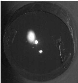
 The system consists of two collimated IR LED channels (for right and left eyes) that illuminate the eye's pupil at an angle of 14 deg, for IOL tilt and decentration measurements, and two double LEDs for phakometry. A CCD camera provided with a telecentric lens placed in front of the eye, conjugated to the pupil captures images of the pupil and Purkinje images. A fixation channel with a minidisplay at a plane conjugate to the retina with a Badal optometer allow projection of visual stimuli, correction of refractive errors and induction of accommodative efforts.
The system is automatically controlled with a program written in Visual Basic and custom routines allow processing of the Purkinje images. Crystalline lens radii of curvature are estimated using equivalent mirror or iterative methods. The lens tilt and decentration is estimated assumming linear relationships between lens, tilt and decentration, using coefficients customized to each subject (obtained from eye models using individual corneal topography and ocular biometry). Tilt of the lens is referred to the pupillary axis and decentration to pupil center.
The system has been used to assess changes of lens radii of curvature with accommodation in humans (in comparison with Scheimpflug imaging) and in rhesus monkeys, and to measure tilt and decentration of the lens in normal eyes and eyes with intraocular lenses.